Figure 1.
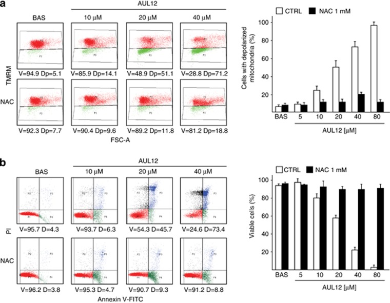
AUL12 induces mitochondrial depolarization and cell death through mitochondrial superoxide production. (a) Cytofluorimetric analysis (Forward Scatter, FSC, versus Tetramethylrhodamine methyl ester, TMRM) showing mitochondria depolarization in human osteosarcoma SAOS-2 cells exposed to AUL12. One representative experiment is reported on the left, in which viable cells (V, TMRM positive) are delimited by the upper quadrant, and cells displaying depolarized mitochondria (Dp) are delimited by the lower quadrant. (b) Death induction on SAOS-2 cells exposed to AUL12 is shown as cytofluorimetric analysis of propidium iodide (PI) versus Annexin V-FITC staining. On the left, one representative experiment is reported. Viable cells (V, double negative for PI and Annexin V-FITC) are delimited by the lower left quadrants; early apoptotic cells (Annexin V-FITC single positive) are in the lower right quadrants; late apoptotic and/or necrotic cells (PI and Annexin V-FITC double positive) are in the upper right quadrants; necrotic cells (PI single positive) are in the upper right quadrants. D (dead) indicates the sum of all apoptotic and necrotic cells. Both in (a) and in (b), data quantification is in the bar graphs on the right; values are the mean±S.D. of at least five experiments. All along the figure, numbers in plots are percentages; AUL12 was incubated for 3 h; N-acetyl cysteine (NAC, 1 mM) was pre-incubated 1 h before AUL12 treatment

