Figure 4.
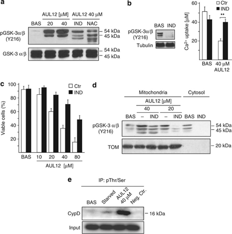
AUL12 induces cells death through ROS-dependent activation of GSK-3α/β. (a) Western immunoblotting analysis showing phosphorylation of the activator Y(279/216) residue of GSK-3α/β in total SAOS-2 cell lysates. Where indicated, cells were treated with AUL12 (2 h) and pre-incubated with NAC (1 mM, 1 h) or with the GSK-3α/β inhibitor indirubin (IND, 2 μM, 3 h) before exposure to AUL12. (b) CRC experiments on whole cells treated or not with AUL12 (40 μM, 2 h) show that pre-treatment with indirubin (IND) rescues PTP opening induced by the chemotherapeutic. Mitochondrial Ca2+ uptake in the various conditions is compared with that measured in unstimulated cells. In the inset, the efficacy of indirubin to inactivate GSK-3α/β is shown as dephosphorylation of the Y(279/216) residue. (c) Death induction on SAOS-2 cells exposed to different concentrations of AUL12 with or without pre-incubation with IND is analyzed by cytofluorimetry with a propidium iodide/Annexin V-FITC double staining, as in Figure 1B. Viable cells reported in the bar graph are negative for both fluorophores. (d) Western immunoblotting analysis of phospho-Y(279/216) GSK-3α/β residue in mitochondrial and cytoplasmic fractions of SAOS-2 cells exposed to AUL12 with or without pre-incubation with IND. (e) Immunoprecipitation of anti-phospho-serine/threonine residues from mitochondria of SAOS-2 cells exposed to serum starvation (24 h) or to AUL12 was followed by Western immunoblotting analysis of cyclophilin D. All along the figure, bar graphs report mean±S.D. values (n=3). Statistical significance was measured with a Student's t test and is indicated by asterisks (**P<0.005). The inhibitor IND (2 μM) was preincubated for 3 h

