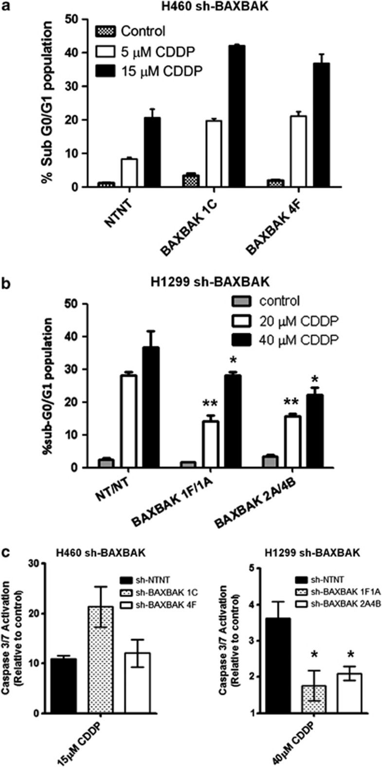
Figure 2.
Cisplatin induces apoptosis in H460 sh-BAXBAK cells but not H1299 sh-BAXBAK cells. (a) Flow cytometry of PI-stained H460 clones after 48 h treatment with cisplatin. Apoptotic cells are indicated by increase in sub-G0/G1 population. (b) Flow cytometry of of PI-stained H1299 clones after 48 h treatment with cisplatin. Apoptotic cells are indicated by increase in sub-G0/G1 population (*P<0.001, **P<0.01). Data are expressed as mean±S.D. (c) Luminescent DEVD-ase assay measuring caspase-3-like activity in sh-NTNT and sh-BAXBAK cells from H460 and H1299 after 24 h treatment with cisplatin, shown relative to untreated controls. Data are expressed as mean±S.D. (*P<0.001)

