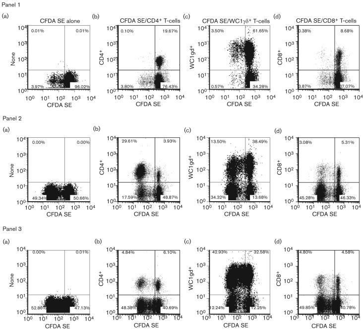
Fig. 3.
Proliferation of T-cell subsets monitored by CFDA SE labelling in response to in vitro restimulation with FMDV vaccine antigen and p252. PBMC from cattle previously vaccinated with O1 Manisa FMD commercial vaccine were labelled with CFDA SE prior to their culture in vitro for 6 days in the presence of medium alone (panel 1), inactivated O1 Manisa FMDV vaccine antigen (panel 2) or FMDV p252 (panel 3). At the end of the culture period, the cells were stained for expression of cell surface differentiation antigens without (a) or with APC-conjugated cc8 (CD4+ T-cells, b), cc15 (WC1+ γδ T-cells, c) and cc63 (CD8+ T-cells, d) mAbs, and analysed by flow cytometry. The percentages of cells in each quadrant are illustrated. One representative dataset (animal FMD 4) from three independent experiments using two animals is shown.

