Fig. 5.

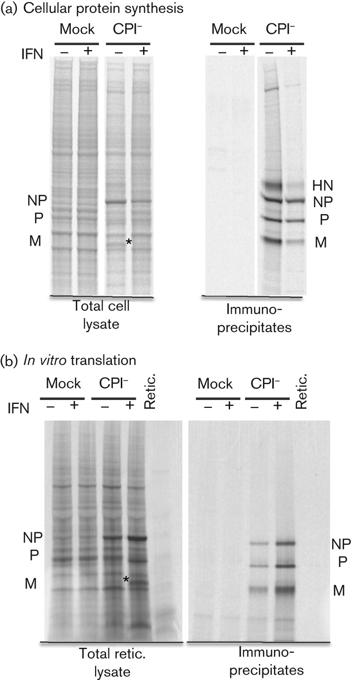
Comparisons of the relative amounts of viral proteins synthesized in infected cells and by in vitro translation of mRNA isolated from infected cells. Hep2/BVDV-Npro cells were mock infected or infected with CPI− at a high m.o.i. and treated or not with IFN at 8 h p.i. (a) At 20 h p.i., the cells were radioactively labelled with [35S]methionine for 1 h and the viral proteins immunoprecipitated. (b) At 20 h p.i., mRNA was isolated from parallel cultures and subject to in vitro translation using a rabbit reticulocyte lysate. Viral proteins were immunoprecipitated or not prior to being separated by electrophoresis through a 7–12 % gradient polyacrylamide gel, and labelled proteins were visualized using a phosphoimager. The position of the M protein in the total cell samples is indicated by asterisks.
