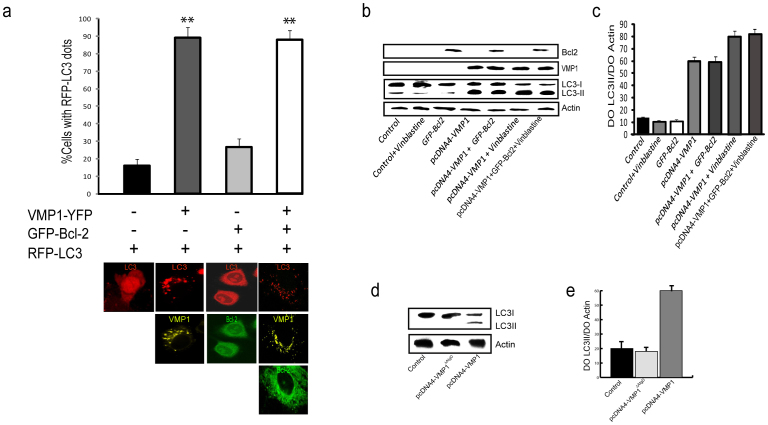
Figure 4. VMP1-Beclin 1 interaction favors autophagosome formation.
(a) HeLa cells co-transfected with RFP-LC3, VMP1-YFP and GFP-Bcl-2. Fluorescence microscopic quantitation of autophagy (% cells with RFP-LC3 dots) was determined as described under “Materials and methods”. Representative images of HeLa cells co-transfected with RFP-LC3, VMP1-YFP and/or GFP-Bcl-2 are shown below graphic bar. (b) Lysates of pcDNA4-VMP1 and GFP-Bcl-2 plasmid-transfected HeLa cells with or without vinblastine treatment were analyzed by western blot. LC3-I (apparent mobility, 18kDa) and LC3-II (16kDa), VMP1, Bcl-2 and Actin proteins were determinated. (c) Quantification of protein signal intensities from western blots showing LC3-II levels after normalization to the control protein Actin. (d) Lysates of pcDNA4-VMP1 and pcDNA4-VMP1ΔAtgD plasmid-transfected HeLa cells were analyzed by western blot. LC3-I (apparent mobility, 18kDa) and LC3-II (16kDa) were determinated. (e) Quantification of protein signal intensities from western blots showing LC3-II levels after normalization to the control protein Actin. Results represent mean ± SD for combined data from four independent experiments. Asterisks indicate a significant difference versus control in two-way ANOVA (**p< 0.01).

