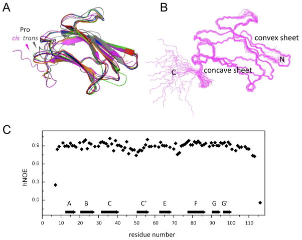
Figure 1.
(A) Superimposition of ribbon structures of N-βGRP from several insects: P. interpunctella - purple, 2KHA (NMR, current work); B. mori - gray, 2RQE [NMR; (11)]; D. melanogaster - green, 3IE4 [X-ray; (13)]; B. mori (laminarihexaose-bound) – orange, 3AQX [X-ray; (12)]; P. interpunctella - blue, 3AQY [X-ray; (12)]; P. interpunctella (laminarihexaose-bound) - red, 3AQZ [X-ray; (12)]; The NMR structure of P. interpunctella N-βGRP reported here differs from that of the B. mori protein (11) in the configuration of a Pro, as indicated. (B) Ensemble of twenty lowest-energy structures of P. interpunctella N-βGRP (2KHA). (C) A plot of backbone heteronuclear {15N}-1H NOE values vs. residue number for P. interpunctella N-βGRP. Pro residues do not give rise to {15N}-1H NOEs. A couple of C-terminal residues had hetero NOE peak intensities barely above the noise level. Each β-strand is represented by an arrow.

