Figure 4.
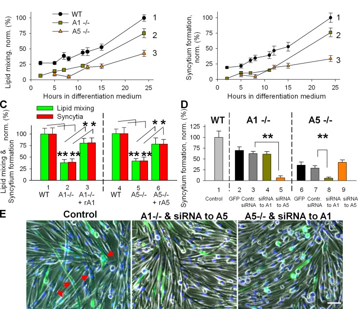
Inhibition of myotube formation for primary myoblasts isolated from either Anx A1−/− or Anx A5−/− mice. (A and B) The lack of either of the two Anxs substantially inhibited lipid mixing (A) and syncytium formation (B) assayed at different times after placement of the cells into DM (B). Curves show myoblasts isolated from WT mice (1), Anx A1−/− mice (2), and Anx A5−/− mice (3). (C) Application of rA1 or rA5 rescues the fusogenic potential of the Anx-deficient myoblasts. Lipid mixing (green) and syncytium formation (red) at 12 h after placing WT myoblasts (1) and Anx A1−/− myoblasts (2) into DM. After 10 h of incubation of Anx A1−/− myoblasts in DM, we applied rA1 (3). Fusion was assayed 2 h later. Fusion in 2 and 3 was normalized to fusion in 1. Cell fusion assayed at 14 h after placement of myoblasts from WT mouse (4) and Anx A5−/− myoblasts (5) into DM. After 12 h of incubation of Anx A5−/− myoblasts in DM, we applied rA5 (6). Fusion was assayed 2 h later. Fusion in 5 and 6 was normalized to fusion in 4. (D) Myoblasts from Anx A1−/− (2–5) or Anx A5−/− (6–9) were cotransfected with GFP vector and with either control siRNA (3 and 7) or siRNA to Anx A1 (4 and 8) or Anx A5 (5 and 9). (2 and 6) Cells transfected only with GFP vector. For each condition, we quantified the efficiency of formation of GFP-labeled syncytia after 24 h in DM and normalized the results to the efficiency of syncytium formation in WT myoblasts (1). (E) Phase-contrast images with nuclear staining (blue) and GFP fluorescence (green) showing WT myoblasts cotransfected with control siRNA and GFP (left), Anx A1−/− myoblasts cotransfected with Anx A5 siRNA and GFP (middle), or Anx A5−/− myoblasts cotransfected with Anx A1 siRNA and GFP (right). All cells are after 24 h in DM. Arrows mark the GFP-labeled multinucleated cells. Bar, 50 µm. (C and D) All results are shown as means ± SEM (n ≥ 3). Levels of significance are shown: **, P < 0.01; *, P < 0.05.

