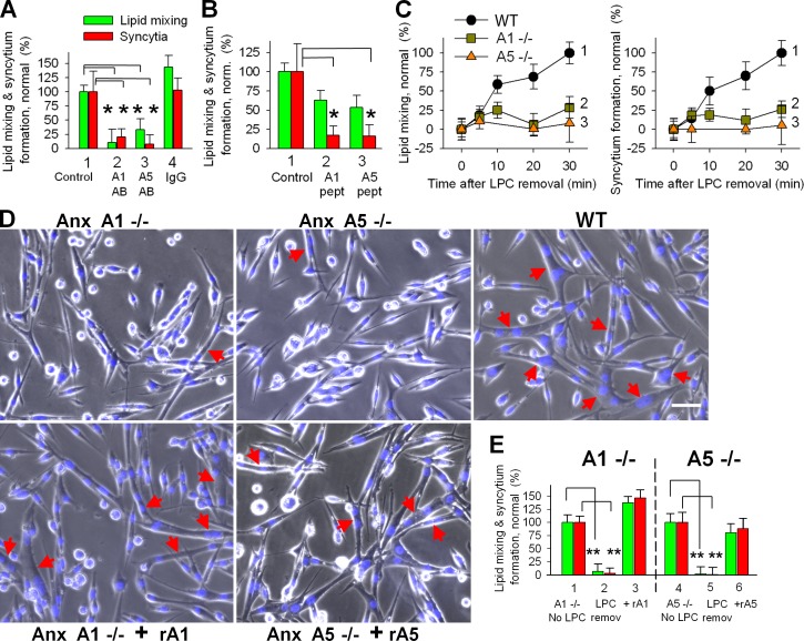
Figure 6.
Synchronized fusion of primary myoblasts is inhibited by antibodies, A1- and A5-peptides, and the lack of either of these Anxs. (A) Antibodies to Anx A1 (2) or A5 (3) or nonspecific IgG (4) were applied at the time of LPC removal. (1) Cells released from LPC block with no immunoglobulins added. (B) A1- and A5-peptides (2 and 3) were applied at the time of LPC removal. (A and B) Lipid mixing (green) and syncytium formation (red) were scored 30 min after LPC removal and normalized to those in the control experiments (1). (C) The extents of lipid mixing (left) and syncytium formation (right) observed at different times after LPC removal at t = 0 for WT, Anx A1−/−, and Anx A5−/− (curves 1,2, 3, respectively) myoblasts. (D) Phase-contrast images with nuclear staining (blue) showing WT, Anx A1−/−, and Anx A5−/− myoblasts 30 min after LPC removal. (bottom) Anx A1−/− myoblasts with rA1 (left) and Anx A5−/− myoblasts with rA5 (right). Anxs were applied at the time of LPC removal. Arrows mark the multinucleated cells. Bar, 50 µm. (E) rA1 and rA5 restore fusogenic potential of Anx-deficient myoblasts. Lipid mixing (green) and syncytium formation (red) in Anx A1−/− (2 and 3) or Anx A5−/− (5 and 6) myoblasts were scored 30 min after LPC removal in the absence (2 and 5) or in the presence of rA1 (3) or rA5 (6). Fusion extents were normalized to those observed for Anx A1−/− (1) and Anx A5−/− (4) myoblasts 24 h after placement of the cells into DM in the experiments in which myogenesis was not interrupted by LPC. All results are shown as means ± SEM (n ≥ 3). Levels of significance relative to corresponding controls (1 in A and B and 1 or 4 in E) are shown: **, P < 0.01; *, P < 0.05.

