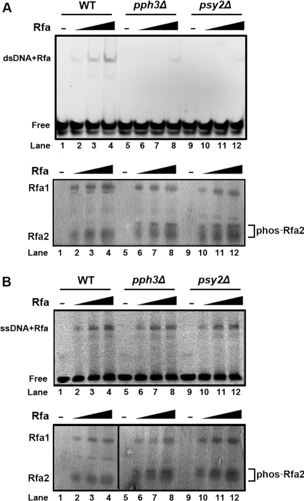
Figure 3. EMSA of Rfa2 with dsDNA and ssDNA.
(A) Top panel: Rfa2 affinity-purified from WT (HT41), pph3Δ (HT42) and psy2Δ (HT43) cells was incubated with FITC-labelled dsDNA at molar ratios of DNA/protein of 0, 8, 16 and 32 at room temperature for 30 min. The protein/DNA mixture was then resolved by TGE/PAGE (8% gel) and detected by fluorescence at an emission wavelength of 320 nm. Bottom panel: the same amounts of purified Rfa2 as above were resolved by SDS/PAGE (12% gel) as a protein loading control. (B) The experiment described above was repeated using ssDNA instead of dsDNA.

