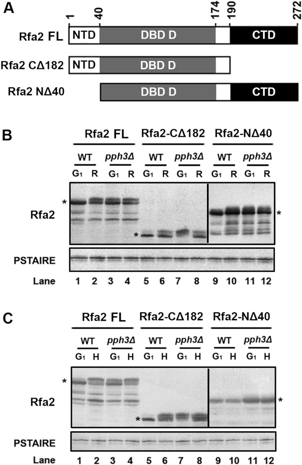
Figure 5. Different domains on Rfa2 are (de)phosphorylated under different circumstances.
(A) Schematic description of FL and truncated versions of Rfa2. Rfa2-FL (HT1 and HT2), Rfa2-CΔ182 (HT31 and HT33) containing residues 1–190 and Rfa2-NΔ40 (HT30 and HT32) containing residues 41–272 were expressed in WT or pph3Δ (SJL2) mutant cells. (B) Western blot analysis of Rfa2 extracted from WT (HT1, HT30 and HT31) and pph3Δ (HT2, HT32 and HT33) cells expressing different versions of Rfa2 in G1-phase and after release. An anti-PSTAIRE antibody was used to detect Cdc28 as a loading control. (C) Western blot analysis of Rfa2 extracted from the same set of cells as above during G1-phase and after HU treatment.

