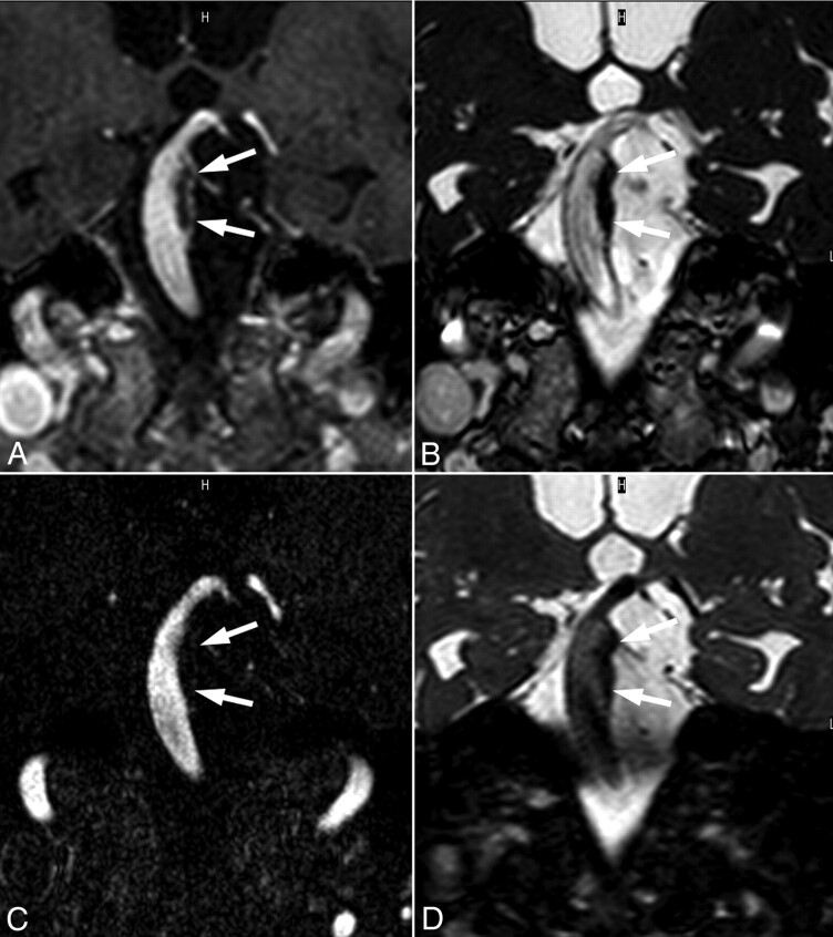
Fig 4.
This partially thrombosed (arrows) basilar aneurysm is demonstrated on postcontrast T1-weighted (A), SSFP (B), CE-MRA (C), and T2-weighted (D) images. These matched oblique coronal scanning planes reveal the extent of thrombosis and further indicate delayed enhancement of the arterial wall underlying the thrombosis (A).

