Figure 1.
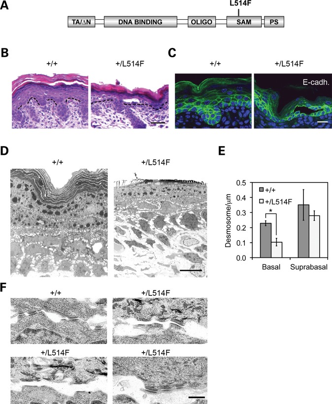
Intra-epidermal blistering and desmosomal defects in AEC mutant mice. (A) Schematic representation of AEC mutant protein generated by knock-in strategy. The L514F mutation falls in the Sterile Alpha Motif (SAM) at the carboxyl-terminus of the p63 alpha isoform. The other p63 domains are also indicated: the TA or the ΔN are two mutually exclusive transactivation domains at the N-terminus of the p63 protein. Oligo: oligomerization domain. PS, post-SAM domain. (B) H&E staining of the skin reveals blistering between the basal and suprabasal compartment in mutant (+/L514F) but not in wild-type (+/+) epidermis. Dashed lines indicate the border between epidermis (top) and dermis (bottom). Scale bar: 50 µm. (C) Immunofluorescence staining for E-cadherin reveals normal localization of this adherens junction component, and confirms basal to suprabasal blistering. Scale bar: 20 µm. (D) Transmission electron microscopy of skin samples reveals reduced basal–basal and basal–suprabasal cell contacts in mutant epidermis. Scale bar: 8 um. (E) Desmosomes between basal–basal and basal–suprabasal cells (basal) are significantly less abundant in mutant (white) versus wild-type epidermis (grey). *P-value = 0.015; n = 6. Desmosomes between suprabasal cells are not significantly affected. (F) Loss of desmosomes in AEC mutant epidermis occurs by disruption of extracellular contacts between adjacent cells, or less frequently by disruption between the desmosome plaque and the intermediate filaments (lower right panel). Scale bar: 500 nm.

