Figure 6.
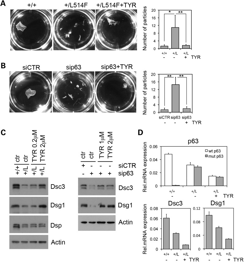
p63 is required for mechanical stress resistance in mouse keratinocytes. (A) AEC mutant and wild-type mouse keratinocytes were cultured in 0.6 mm calcium for 24 h, either in the presence or in the absence of 2 mm Tyrphostin (TYR), and then treated with dispase. Detached monolayers were subjected to mechanical stress, and resulting fragments of the cell sheet were imaged and counted. Quantitative evaluation of particles generated in the experiment is shown on the right (*P-value <0.05, **P-value <0.01; n = 10). Error bars denote SD. (B) p63-depleted (sip63) or control (siCTR) keratinocytes were treated as in (A). (**P-value <0.01; n = 10). Error bars denote SD. (C) Immunoblotting of desmosomal proteins in total cell extracts of wild-type (+/+) and AEC mutant (+/L) primary keratinocytes (left panels) or control (siCTR) and p63 knockdown keratinocytes (sip63) (right panels) were cultured in 0.6 mm calcium for 24 h and either untreated (ctr) or treated with AG1478 (TYR) at the indicated concentrations (left panel) 2 h before calcium addition. (D) Real-time RT–PCR on RNA isolated from wild-type (+/+) and mutant keratinocytes (+/L). Mutant (grey bars) and wild-type p63 (white bars), Dsc3 and Dsg1 mRNA were measured in keratinocytes treated as in (C). TYR was used at 2 µm. Data are normalized for actin mRNA levels and are represented as mean +/− SD normalized mRNA levels.

