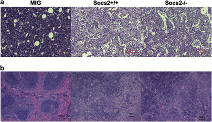
Figure 4.
Histopathologic staining of BM and spleen after disease onset. (a) Hematoxylin eosin staining of BM sections showing increased granulopoiesis and enlarged sinusoids both in mice transplanted with Socs2+/+ and Socs2−/− cells compared with MIG. (b) Hematoxylin eosin staining of spleen with marked pathology, including severe infiltration of hematopoietic cells at various maturation stages, after Socs2+/+ and Socs2−/− transplants.

