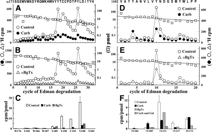
Figure 3.
[3H]Physostigmine photolabeling at the α-γ extracellular interface in the presence or absence of Carb or α-BgTx. A, B, D, E, 3H (○, ●, ▵) and PTH-amino acids (□) released during sequence analysis of fragments beginning at αSer-173 (A, B) and γVal-102 (D, E) isolated from Torpedo nAChR photolabeled with [3H]physostigmine in the absence of other drugs (A, B, D, E, ○, □), in the presence of 1 mm Carb (A, D, ●), or in the presence of 8 μm α-BgTx (B, E, ▵). C, F, Quantification of [3H]physostigmine photoincorporation (in cpm/pmol) into aromatic amino acid residues within the primary structure of fragment beginning at αSer-173 (C) or γVal-102 (F). For the control and +Carb conditions, the values plotted are the mean ± SD from two independent photolabeling experiments, whereas data for +α-BgTx are from a single experiment. To calculate the efficiency of labeling at αCys-192 (cycle 20) from sequencing data shown in A, the background 3H release in cycle 20 originating from the photolabeling of αTyr-190 in cycle 18 was estimated by fitting the observed 3H releases in cycles 18, 19, and 25 to an exponential decay. F, Also included are values from a third [3H]physostigmine preparative photolabeling in the presence of Carb or Carb and galanthamine and the efficiency of photolabeling of δTyr-172, derived from sequencing data presented in Figure 4.

