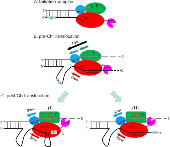
FIGURE 6.
Schematic of RecBCD-DNA complexes pre- and post-CHI. A, RecBCD-DNA initiation complex. RecD (green) and the secondary RecBC translocation site (blue) operate on the 5′-ssDNA tail, and the primary RecBC translocase site (red) operates on the 3′-ssDNA tail. B, pre-CHI DNA unwinding by RecBCD. The 5′ to 3′ rate is faster due to the action of two translocases (RecD (green) and the secondary RecBC (blue)) versus only one translocase (primary RecBC) acting in the 3′ to 5′ direction, resulting in a loop in the 3′-terminated strand ahead of the enzyme. C, post-CHI DNA unwinding by RecBCD. Upon deactivation of RecD, the 3′ to 5′ and 5′ to 3′ translocation rates are equal due to the concerted action of the RecBC secondary (5′ to 3′) (blue) and primary (3′ to 5′) (red) translocases, resulting in maintenance of the loop formed in the 3′-terminated strand ahead of the enzyme. Two alternative pathways (I versus II) result depending on whether CHI remains bound to RecC (I) wherein a second loop would form at the interface between RecB and RecC as proposed (13, 41). Alternatively, if the 3′-tail does not remain bound to RecC (II), then the 3′-ssDNA will pass through RecC, and a second loop would not form.

