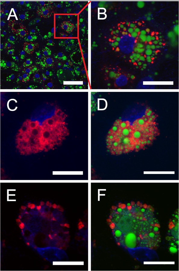
FIGURE 1.
Endodermal epithelial cells take up yVLDL particles. Explants from YS were transferred to Lab Tek Permanox 4-well imaging chambers. After 48 h of EEC outgrowth, the medium was changed to serum-free conditions, and 50 μg/ml DiI-labeled yVLDL (red fluorescence) was added. In A and B, the EECs were incubated with the labeled lipoprotein for 4 h at 37 °C; B shows the magnification of a cell from A. In C–F, the cells were incubated with 50 μg/ml DiI-yVLDL for 4 h, the medium was removed and replaced with medium without DiI-yVLDL, and cells were further incubated for 20 h. In E and F, the incubation medium contained 200 μm chloroquine, an inhibitor of lysosomal degradation. After the incubations, the cells were washed two times with PBS, fixed in 4% PFA in PBS, pH 7.4, for 30 min at room temperature, and washed two times with PBS. After counterstaining with BODIPY 493/503 (Molecular Probes) for the visualization of lipid droplets (green fluorescence) and DAPI for the nuclei, respectively, cells were mounted in Fluorescent Mounting Medium (Dako). Images were obtained with a Zeiss LSM510 confocal microscope. Scale bar, 50 μm (A) or 20 μm (B–F).

