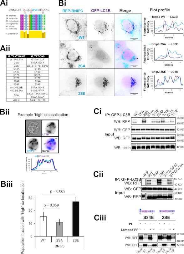
FIGURE 3.
Serine phosphorylation of the Bnip3 LIR motif determines its affinity for LC3B. A, putative serine phosphorylation sites of interest and generated mutants. Ai, alignment of the region containing the Bnip3 LIR across mammalian species. Aii, table describing Bnip3 mutants used in this study. B, phosphorylation state of the LIR determines autophagosomal sequestration of mitochondria. Bi, representative high resolution images of GFP-LC3B colocalization with RFP-Bnip3 WT and the indicated multisite LIR mutants at mitochondria in HeLa cells at 24 h. Plot profiles illustrate colocalization between Bnip3-targeted mitochondria and autophagosomes. Bii, MIFC analysis of Bnip3 WT, 2SA, and 2SE induction of mitophagy. The indicated Bnip3 constructs were expressed with GFP-LC3B in cardiac HL-1 cells for 24 h and imaged using the ImageStream X flow cytometer. Representative images and plot profiles are shown. Biii, using MIFC, quantification of the subpopulation of Bnip3 WT-targeted mitochondria with high GFP-LC3B colocalization was achieved using the colocalization function and gating on the fraction of cells with high colocalization. The identical analysis was applied to Bnip3 mutant populations. Population fractions are indicated as well as the percentage change of the two mutants compared with the wild type. C, localization of serine residues, which control binding affinity with LC3B. Ci, the indicated RFP-Bnip3 WT and multisite and single site mutant constructs were expressed in MCF-7 cells stably expressing GFP-LC3B. At 48 h of expression, immunoprecipitations (IP) were performed with α-GFP. Shown is Western blot detection of RFP, GFP, and actin. Cii, double mutant S17A/S24E and S17E/S24A Bnip3 constructs were expressed in MCF-7 stably expressing GFP-LC3B. At 48 h of expression, immunoprecipitations were performed with α-GFP. Shown are Western blot detection of RFP and GFP. Ciii, in vitro analysis of serine 17 phosphorylation during LC3B binding to Bnip3. Bnip3 S24E and 2SE mutants were expressed for 48 h in MCF-7 cells stably expressing GFP-LC3B. Cell lysates were incubated with λ-phosphatase (800 units) prior to immunoprecipitation with α-GFP. Shown is Western blot detection of RFP and GFP. PI, phosphatase inhibitor.

