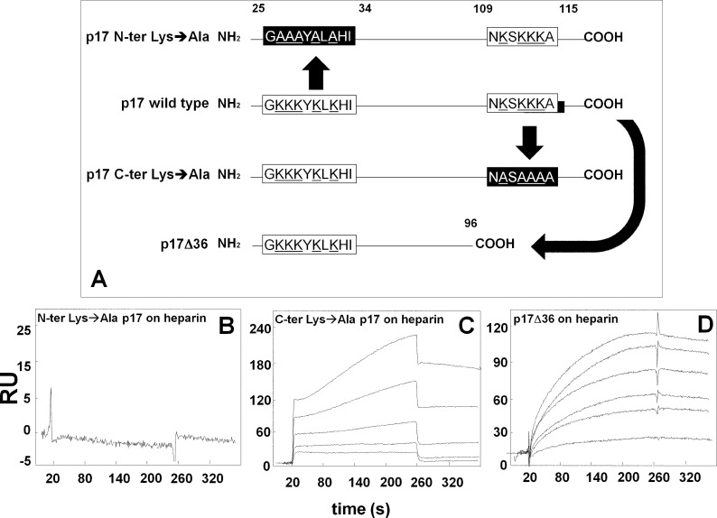
FIGURE 3.
SPR analysis of the interaction of p17 mutants with heparin. A, schematic representation of the p17 mutants used in the present work. The N- and C-terminal basic motifs of p17 (white boxes) were deleted or neutralized (black boxes) by substituting positively charged lysine with alanine (underlined). Blank-subtracted sensorgrams showing the binding of p17 N-terminal (N-ter) Lys→Ala (500 nm) (B), p17 C-terminal (C-ter) Lys→Ala (500, 250, 125, 62.5, and 31.25 nm) (C) and p17Δ36 (950, 900, 800, 500, 250, and 100 nm) (D) to a heparin-coated sensorchip. The response (in RU) was recorded as a function of time.

