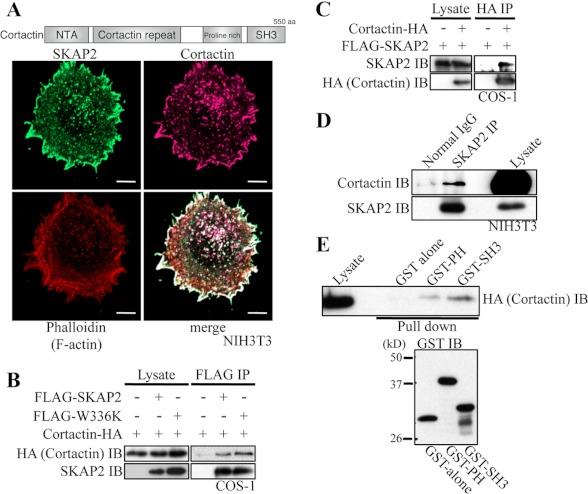
FIGURE 5.
SKAP2 interacts with cortactin. A, top, structure of human cortactin. NTA, amino-terminal acidic region; aa, amino acids. Bottom, NIH3T3 cells were immunostained using anti-SKAP2 (green) and anti-cortactin (magenta) antibodies and phalloidin (red). Bar, 10 μm. B, COS-1 cells were transfected with cortactin-HA with or without FLAG-SKAP2 or W336K mutant. FLAG-SKAP2 was immunoprecipitated using an anti-FLAG antibody. Cortactin and SKAP2 were detected using anti-HA and anti-SKAP2 antibodies, respectively. C, COS-1 cells were transfected as indicated. Cortactin-HA was immunoprecipitated (IP) using an anti-HA antibody, and SKAP2 was detected. D, endogenous SKAP2 in NIH3T3 cells was immunoprecipitated using an anti-SKAP2 antibody. Cortactin and SKAP2 were detected using anti-cortactin and anti-SKAP2 antibodies, respectively. E, top, protein extracts from COS-1 cells transfected with cortactin-HA were pulled down by GST alone, the GST-PH domain, or the SH3 domain of SKAP2 and immunoblotted (IB) with an anti-HA antibody. Bottom, immunoblot of GST fusion proteins was shown.

