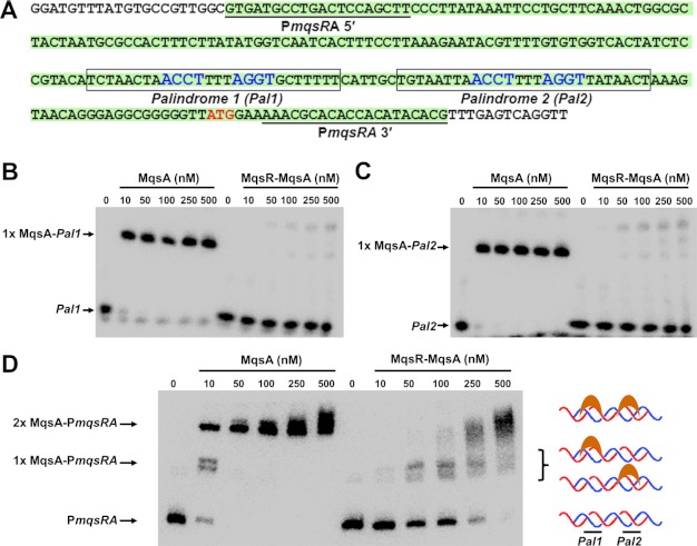
FIGURE 3.
The MqsR-MqsA complex does not bind to the mqsRA promoter in vitro. A, sequence of the mqsRA promoter. DNA constructs used in this study are illustrated. Palindromes 1 and 2 are boxed (nucleotides that interact specifically with MqsA, as determined from the MqsA-DNA crystal structure, are in blue). The PmqsRA DNA is highlighted in green, and the primers used to amplify it are underlined. B, EMSA with increasing amounts of either MqsA alone (lanes 2–6) or the untagged MqsR-MqsA complex (lanes 8–12) incubated with biotin-labeled palindrome 1 (Pal1) of PmqsRA. C, same as B, except proteins were incubated with biotin-labeled Palindrome 2 (Pal2). D, same as B, except proteins were incubated with biotin-labeled PmqsRA promoter DNA. The two shifted bands represent protein bound to either one (middle arrow; 1× MqsA-PmqsRA) or both palindromes (top arrow; 2× MqsA-PmqsRA). DNA binding in the presence of the MqsR-MqsA complex is due to trace amounts of free MqsA, as the observed migration positions are identical to that seen with MqsA alone. For all gels, the negative control (100 fmol of labeled DNA lacking protein) is shown in lanes indicated as 0.

