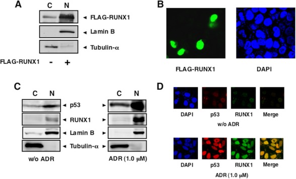
FIGURE 3.
Nuclear accumulation of RUNX1 upon ADR treatment. A and B, nuclear accumulation of the exogenous RUNX1. HCT116 cells were transiently transfected with FLAG-RUNX1 expression plasmid. Forty eight hours after transfection, cells were fractionated into cytoplasmic (C) and nuclear (N) fractions. These fractions were analyzed by immunoblotting with anti-FLAG antibody. The amounts of tubulin-α and lamin B were also examined as a cytoplasmic nuclear and a nuclear marker, respectively (A). For immunostaining, HCT116 cells were transiently transfected with the expression plasmid encoding FLAG-RUNX1. Forty eight hours after transfection, cells were fixed and incubated with anti-FLAG antibody (green). Cell nuclei were stained with DAPI (blue) (B). C and D, ADR-mediated nuclear accumulation of the endogenous RUNX1. HCT116 cells were treated with 1 μm ADR or left untreated. Twenty four hours after ADR treatment, cells were fractionated as in A, and each fraction was analyzed by immunoblotting with the indicated antibodies (C). For immunostaining, HCT116 cells were treated with 1 μm ADR or left untreated. Twenty four hours after ADR exposure, cells were simultaneously incubated with anti-p53 (red) and anti-RUNX1 (green) antibodies. The merged image (yellow) shows the nuclear co-localization of RUNX1 and p53. Cell nuclei were stained with DAPI (blue) (D). w/o, without.

