FIGURE 1.
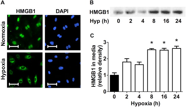
Hypoxia induces HMGB1 release in HPAECs. A, HPAECs exposed to 8 h of hypoxia (1%) were assessed for HMGB1 release by immunofluorescent staining (HPAECs, green; DAPI, blue; scale bars, 25 μm). Photomicrographs are representative of three independent experiments. B and C, Western blot analysis (B) and quantitative densitometry (C) of HMGB1 accumulation in the cell media of HPAECs exposed to hypoxia for the indicated times are shown. Blots are representative of four independent experiments. *, p < 0.05; error bars, S.E.

