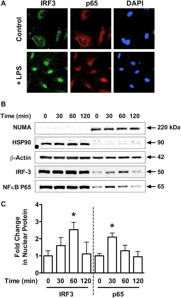
FIGURE 7.
LPS stimulates MyD88-dependent and -independent TLR4 signaling in HPAECs. A, HPAECs exposed for 1 h to LPS (1 μg/ml) were assessed for IRF3 nuclear translocation by immunofluorescent staining against IRF3 (green; nuclear stain DAPI, blue; scale bar, 25 μm). Images are representative of three independent experiments. B, Western blot analysis is shown of NUMA (150 kDa, nuclear marker), HSP90 (90 kDa, cytoplasmic marker), β-actin (42 kDa), NFκB-p65 (65 kDa), and IRF3 (50 kDa) in cytoplasmic and nuclear fractions from HPAECs treated with LPS (1 μg/ml) for the indicated times. Blots are representative of three independent experiments and are quantified in C. Data represent the mean ± S.E. (error bars) of three independent experiments. Analysis of variance; *, p < 0.05 versus unstimulated control.

