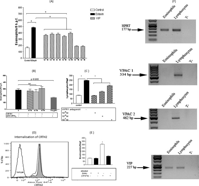
FIGURE 3.
A, VIP eosinophilotactic activity compared with eotaxin. Results are ± S.E. of 10 independent experiments performed in triplicate. *, p < 0.05. B, modulatory effects of VIP-R1 and anti-CRTH2 blocking antibody on 10−7 m VIP eosinophilotactic activity. Results are ± S.E. (error bars) of six independent experiments performed in triplicate. The culture medium consisted of a solution of Hanks' balanced salt solution for all treatments (buffer only, VIP-R1, and anti-CRTH2) during the chemotaxis assays. C, blocking effect of VIP-R1 on 10−7 m VIP-induced lymphocyte chemotaxis. Results are the mean ± S.E. (error bars) of five independent experiments performed in triplicate. *, p < 0.05. D, internalization of surface CRTH2 receptors in human eosinophils after a 5-min stimulation with 10−7 m PGD2. The gray histogram represents CRTH2 spontaneous expression, whereas the white histogram represents CRTH2 expression after stimulation with 10−7 m PGD2 for 5 min. E, effect of the treatment of eosinophils with 10−7 m PGD2, 5 min before and during the chemotaxis assay, against 10−7 m VIP (mean ± S.E. (error bars) of five independent experiments performed in triplicate). White bars represent migration in the absence of PGD2, whereas black bars represent migration in the presence of PGD2. F, RT-PCR showing expression of VPAC1 and VPAC2 by human lymphocytes but not human eosinophils, whereas VIP protein was expressed by both cell types.

