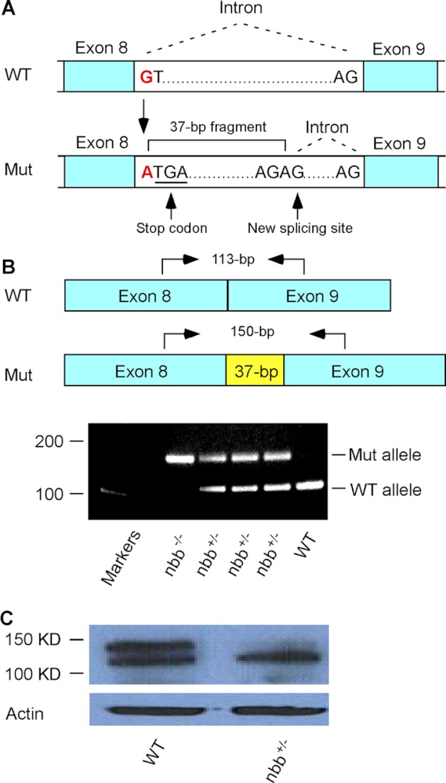
FIGURE 2.
Analysis of zebrafish nbb mutation. A, a diagram that shows the mutation at the beginning of intron 8–9. The substitution of nucleotides from G to A altered the donor splicing site, resulting in a 37-bp insertion with a premature stop codon. B, RT-PCR analysis of Stil expression in wild-type, heterozygous, and homozygous nbb mutants. In the diagram, arrows indicate RT-PCR primer sites used to amplify the region at exon 8–9 junction from cDNA. In heterozygous mutants (nbb+/−), both wild-type (113-bp) and mutant (150-bp) alleles were amplified. In homozygous mutants (nbb−/−), only the 150-bp fragment was amplified. In wild-type embryos, the 113-bp fragment was amplified. C, Western blot of retinal lysates from wild-type and heterozygous nbb mutant fish. The mutant fish showed decreased STIL protein expression. Mut, mutant.

