FIGURE 4.
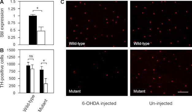
Toxicity of DA-IPCs to 6-OHDA in wild-type and mutant retinas. A, relative Stil mRNA expression in wild-type and nbb mutant retinas. The expression of Stil in wild-type retinas was normalized to 1 (black bar). Note the decrease of Stil expression in nbb mutant retinas (white bar). Data represent the means ± S.E. (n = 4 in each group). *, p < 0.01. B, number of anti-TH positive retinal DA-IPCs after injection of subtoxic 6-OHDA (white bars) or PBS (black bars). In wild-type fish, injection of subtoxic 6-OHDA produced no effects on DA-IPCs. In nbb retinas, the same treatment resulted in degeneration of DA-IPCs. Data represent the means ± S.E. (n = 3 in each group). ns, not significant; *, p < 0.01. C, fluorescent images of flat-mount wild-type and nbb mutant retinas labeled with anti-TH antibodies. Note the decreased number of DA-IPCs in mutant retinas after subtoxic 6-OHDA treatment. The images were taken at the nasal retina adjacent to the optic nerve. Scale bar, 100 μm.

