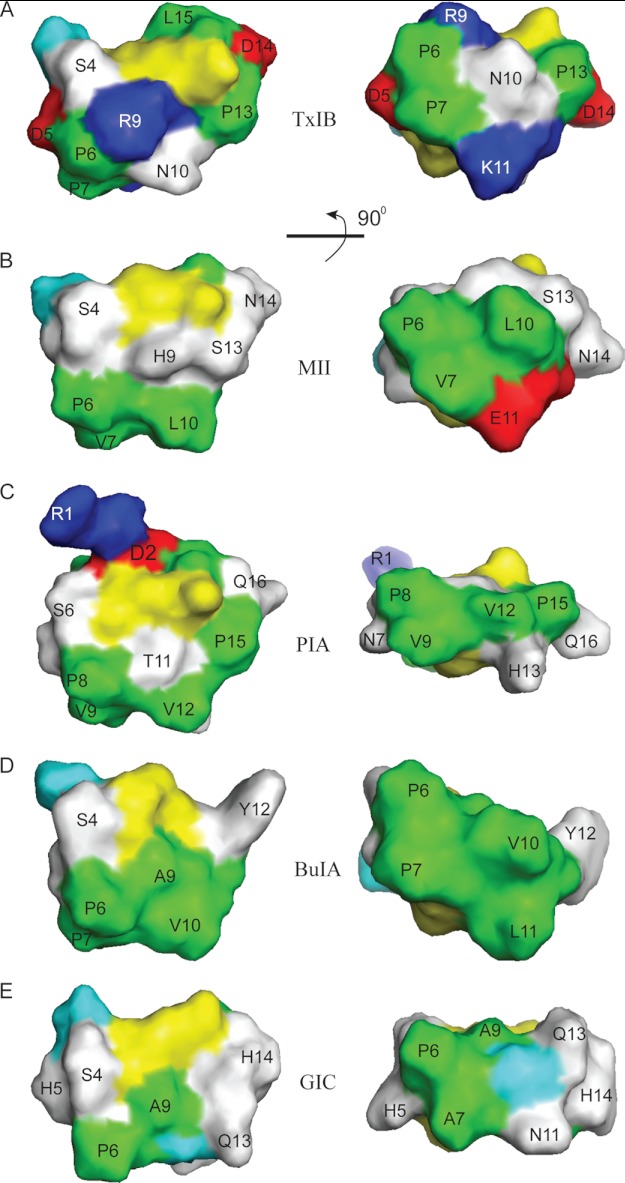
FIGURE 6.
Surface representation of selected α-conotoxins. A, α-CTx TxIB (PDB code 2LZ5); B, α-CTx PIA (PDB code 1ZLC); C, α-CTx BuIA (PDB code 2I28); D, α-CTx GIC (PDB code 1UL2); and E, α-CTx MII (PDB code 1MII). Positively charged residues (Arg and Lys) are shown in blue, negative residues (Asp and Glu) are red, polar residues (Asn, Gln, His, Ser, Thr and Tyr) are white, and hydrophobic residues (Ala, Ile, Leu, Pro, and Val) are green, cystines are yellow, and glycines are cyan. The surface images of the peptides on the right are the images after the 90° rotation of the left images around the horizontal axis.

