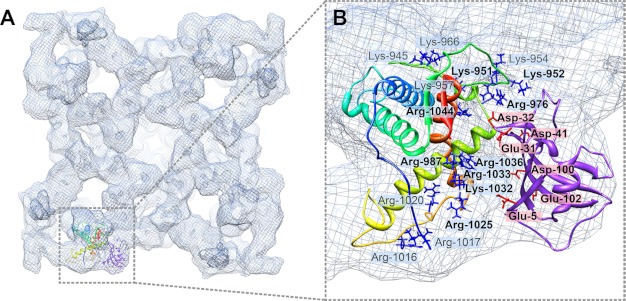
FIGURE 5.
Interaction between modeled structure RyR1(850–1,056) and FKBP12. A, flexibly fitted RyR1 fragment model and rigid-body fitted FKBP12 in the closed conformation RyR1 cryo-EM map, with docked structures shown in only one RyR1 subunit for clarity. B, close-up view of interface between the RyR1 fragment and FKBP12. Positively charged residues in RyR1 are depicted as blue stick structures, and negatively charged residues in FKBP12 are indicated as red stick structures. Residues with numbers in boldface are located at the direct interface. Residue Arg-976 in RyR1 is close enough to residue Asp-32 in FKBP12 to form an intermolecular salt bridge.

