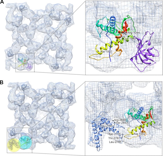
FIGURE 6.
Mapping disease-causing mutations in RyR1 fragment model. A, in the flexibly fitted RyR1 fragment model, mutant residues are depicted as black stick structures. Two residues, Cys-1040 and Arg-1044, are located in helix 4 (orange-red), which is at RyR1/FKBP12 interface, and possibly involved in the RyR-FKBP interaction. B, one residue Ala-1002 is located at interface between domain 9 (cyan sphere) and domain 10 (yellow sphere), where the RyR1 fragment interacts with docked crystal structure 3RQR (residues 2,733–2,940), which is a phosphorylation domain in RyR1. Seven mutant residues in this phosphorylation domain that show direct contacts with the RyR1 fragment model are highlighted with asterisks (in the unstructured loop) or black stick structures.

