FIGURE 2.
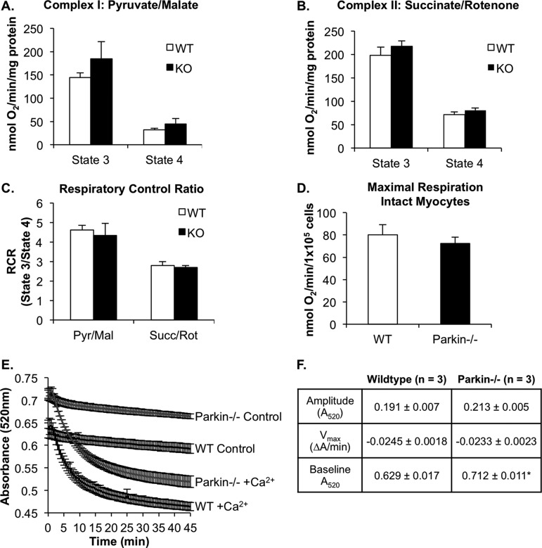
Mitochondrial respiration is normal in Parkin−/− mouse hearts at 3 months of age. A and B, state 3 and state 4 respiration rates of mitochondria isolated from WT or Parkin−/− mouse hearts with substrates for complex I (pyruvate/malate) (A) or complex II (succinate/rotenone) (B). C, respiratory control (RCR) ratios for complex I and complex II substrates (n = 4). Pyr/Mal, pyruvate/malate; Succ/Rot, rotenone/succinate. D, maximal mitochondrial respiration rates in isolated intact WT and Parkin−/− adult mouse myocytes (n = 3). Mean ± S.E. No significant differences were observed. E, swelling of isolated mitochondria in the presence of 150 μm calcium (n = 3). F, the degree and rate of swelling (amplitude and Vmax, respectively) were not significantly different between WT and Parkin−/− mitochondria. Base-line absorbance values for Parkin−/− mitochondria were significantly higher than WT. Mean ± S.E. (n = 3, *, p < 0.05 versus WT).

