
FIGURE 3.
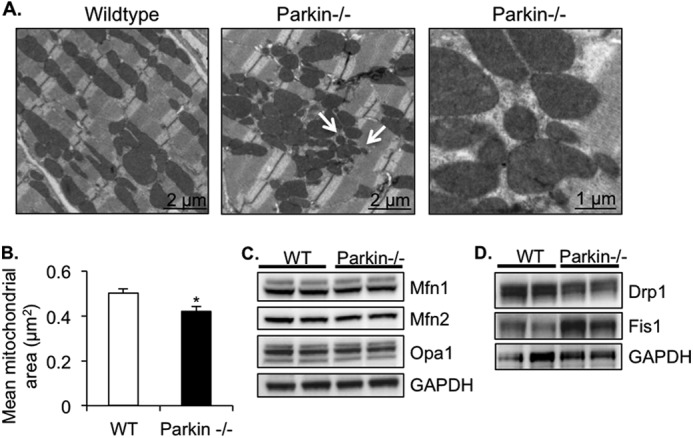
Parkin-deficient hearts have disorganized and smaller mitochondria. A, representative transmission electron micrographs of heart sections from 3-month-old mice. Arrows signify smaller mitochondria. B, quantitation of mean mitochondrial area ± S.E. in WT and Parkin−/− hearts (*, p < 0.05 versus WT). C, Western blot of mitochondrial fusion proteins Mfn1, Mfn2, and Opa1. D, Western blot of mitochondrial fission proteins Drp1 and Fis1.