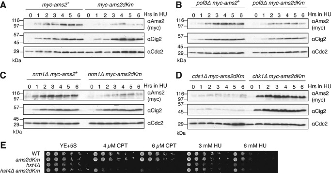
FIGURE 6.
A feedback mechanism of Ams2 levels in G1. A, WT myc-ams2+ (HYY908) and myc-ams2-dKm (HYY909) cells were grown to mid-log phase in YE+5S medium, and hydroxyurea was added to 20 mm final concentration. The samples were taken every hour for analysis by immunoblotting with indicated antibodies. B–D, same as A, but pof3Δmyc-ams2+ (HYY1094) and pof3Δmyc-ams2-dKm (HYY1067) (B), nrm1Δmyc-ams2+ (HYY1117) and nrm1Δmyc-ams2-dKm (HYY1119) (C), or cds1Δ myc-ams2-dKm (HYY1083) and chk1Δ myc-ams2-dKm (HYY1080) (D) cells were analyzed. E, asynchronous WT ams2+ (HYY908), ams2-dKm (HYY909), hst4Δ (HYY1032), and hst4Δams2-dKm (HYY1051) cells were spotted with decreasing cell number onto rich medium containing indicated DNA-damaging agents and incubated for 4 days.

