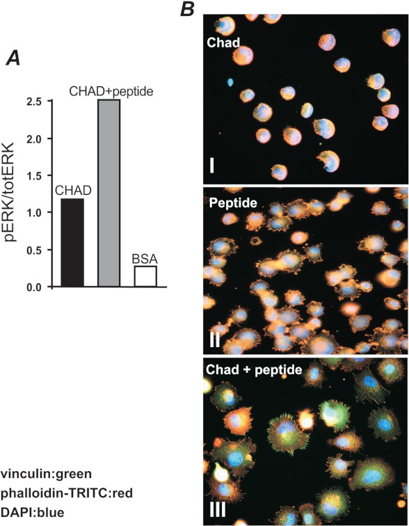
FIGURE 9.
Activation of signaling pathways and rearrangement of cytoskeletal elements upon exposure of cells to the hbd-CKFPTKRSKKAGRH359. A, tyrosine phosphorylation of signaling pathways in cells bound to chondroadherin upon the addition of the hbd-CKFPTKRSKKAGRH359. Tissue culture 6-well dishes (Costar) were coated overnight with 5 μg/ml chondroadherin. The dishes were blocked for nonspecific binding with 0.5% BSA. To determine ERK phosphorylation, primary human chondrocytes were serum-starved and analyzed as described in the legend for Fig. 8. pERK, phospho-ERK; totERK, total ERK. B, immunochemical staining of adhesion complexes. Chamber slides were coated with 5 μg/ml CHAD (top and bottom panels) or 33 μg/ml (20 μm) of the hbd-CKFPTKRSKKAGRH359 peptide (middle panel) and blocked for nonspecific binding with BSA (0.5%). The cells (105kc) were allowed to adhere for 1 h in the absence (top and middle panel) or in the presence (bottom panel) of the peptide (150 μm, 250 μg/ml), and adhesion complexes were identified using antibodies against phalloidin (red) and vinculin (green).

