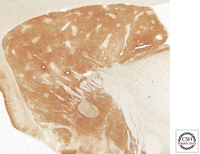
Figure 3.
Striatal compartments. Although the striatum appears as a rather homogeneous structure, several histochemical and immunohistochemical stains evidence the presence of two compartments named striosomes and matrix. The photomicrograph is taken from a parasagittal section through the primate striatum. The immunohistochemical detection of the calcium-binding protein calbindin reveals the presence of a number of patchy areas with weak calbindin stain (striosomes) immersed within a background showing higher calbindin stain (matrix).

