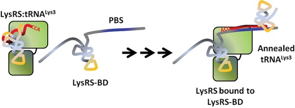
FIGURE 6.

Hypothetical model for TLE-assisted tRNALys3 primer placement. The tRNALys3 primer shares 18 nt of complementarity (red) to the HIV-1 vRNA and must be annealed for reverse transcription to begin from the primer’s CCA-3′OH end. Human LysRS and tRNALys3 are packaged as a complex along with genomic RNA, which has a TLE (yellow) upstream of the primer binding site (PBS) (blue). Although the full extent to which the TLE-containing viral RNA mimics the structure of a tRNA is unknown, the TLE is part of a LysRS binding domain (LysRS-BD) that effectively competes for binding to hLysRS (green, two domains representing catalytic and anti-codon-binding domains). This competition may facilitate release of bound tRNALys3 from the synthetase, which can then be annealed to the vRNA. Arrows indicate intermediate steps in the annealing pathway, which requires viral proteins that facilitate tRNALys3 annealing such as Gag and GagPol (omitted for clarity).
