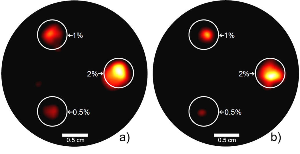
Figure 4.
Reconstructed XFCT images for the 3 cm-diameter phantom. (a) tin-filtered polychromatic beam, 1 minute exposure per projection. (b) lead-filtered polychromatic beam, 3 minute exposure per projection. Each phantom is labeled with the GNP concentration (by weight) of each GNP-loaded column included. The GNP-loaded regions are well delineated, and the tin-filtered beam was able to reconstruct an image using much less scan time and dose. The display window in both figures is 10%–100% of the maximum intensity value.

