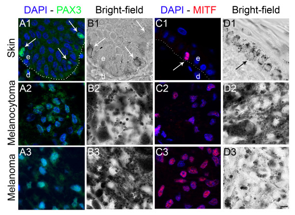
Figure 1.
PAX3 and MITF immunolabeling in horse skin and cutaneous melanocytic proliferations. (1) control horse skin, (2) cutaneous melanocytoma, (3) cutaneous melanoma (A): PAX3 protein immunolabeling (green) with corresponding bright-field photographs (B). A specific nuclear PAX3 labeling is identified in melanocytes (arrows) and melanocytic cells (A1-A3) with low background signal. (C): MITF protein immunolabeling (red) with corresponding bright-field photographs (D). A specific nuclear MITF labeling is observed in melanocytic cells in control skin and lesions (C1-C3) with very low background. Nuclear counterstaining is shown in blue. Dotted lines (A1 and C1) indicate epidermis-dermis boundary. e, epidermis; d, dermis. Magnification is the same in all images, bar: 10 μm.

