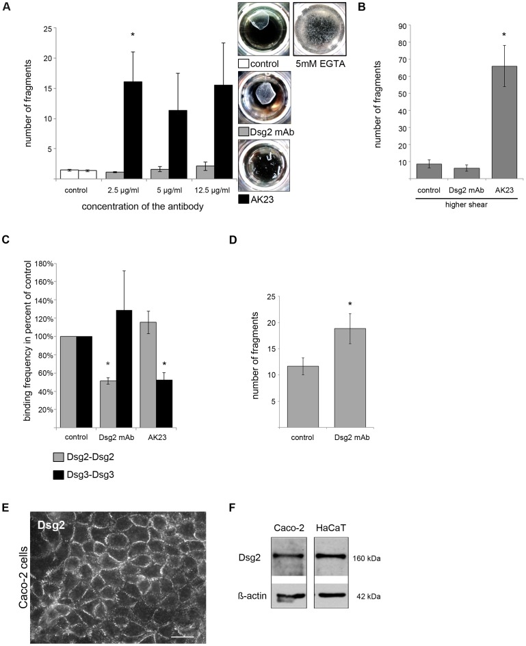
Figure 2. Antibody-mediated targeting of Dsg3, but not of Dsg2, led to profound loss of cell cohesion.
(A) After antibody incubation for 24 h with either a monoclonal Dsg2 antibody (Dsg2 mAb) or AK23, confluent HaCaT monolayers were subjected to dispase-based dissociation assays. Loss of cell cohesion was detectable in cells incubated with AK23 only. (n = 8; * p<0.05 vs. control) Photos were taken immediately after assay performance. 1 hour incubation with 5 mM EGTA led to profound loss of cell cohesion indicating the non-hyperadhesive state of HaCaT cells used for these experiments. (n = 6) (B) Exposing the cells to more severe mechanical stress in dissociation assays increased fragment numbers after AK23 treatment only. (n = 6; * p<0.05 vs. control) (C) Atomic force microscopy was used to demonstrate antibody-mediated interference with homophilic Dsg2 and Dsg3 binding. Both antibodies reduced the binding frequency of their respective antigens. (>1000 force distance cycles on more than 2 different cantilever/substrate combinations; * p<0.05 vs. control) (D) Dispase-based dissociation assay performed with confluent Caco-2 cells after 24 h incubation with Dsg2 mAb showed a significant increase in fragment numbers compared to control cells. (n≥20; * p<0.05 vs. control) (E) Protein expression of Dsg2 in Caco-2 cells was proven by immunofluorescence (scale bar, 20 µm) and (F) Western blot analysis. ß-actin was used as loading control.

