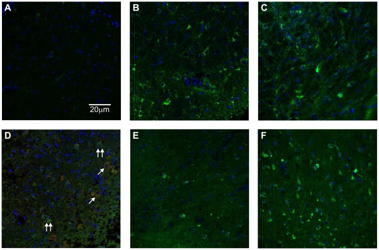
Figure 6. 6h sleep deprivation causes an increase in iNOS expression in BFB and cortex.
iNOS Immunofluorescence (green) was largely absent in BFB slices from rats not sleep deprived after 1 h post-sacrifice incubation in aCSF (a), but strong after 6 h SD with 1 h (b) and 4 h (c) post-sacrifice incubation. The blue immunfluorescence is DAPI, showing nuclei. Double immunfluorescence staining for iNOS and ChaT (red) in BFB for a 6 h SD rat after 1 h incubation is shown in (d), single arrows indicate examples of somata with colocalised ChaT and iNOS, double arrows somata with iNOS but no ChaT. In the cortex, iNOS immunfluorescence after 4 h incubation was present in non-SD rats (e), but less strong than those after 6 h SD (f).

