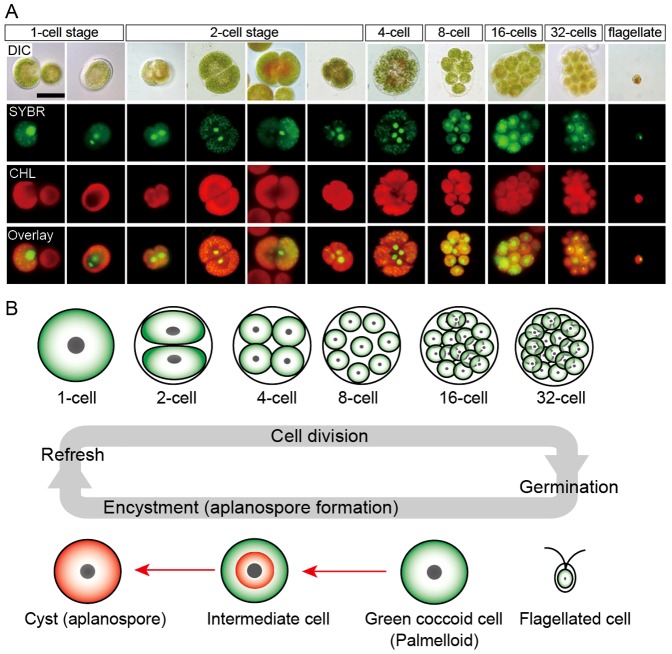
Figure 1. Life cycle of H. pluvialis.
A. Fluorescence microscopy images, showing the 1- to 32-cell stages, and the flagellated stage. DIC: differential interference contrast image; SYBR: SYBR Green I-stained cells (green); CHL: chlorophyll autofluorescence (red); and Overlay: overlaid images of SYBR and CHL. B. Illustration of life cycle of H. pluvialis. Refresh: when old cultures are transplanted into fresh medium, coccoid cells undergo cell division to form flagellated cells within the mother cell wall. Germination: Flagellated cells settle and become coccoid cells. Continuous and/or strong light accelerate the accumulation of astaxanthin during encystment (red arrows).

