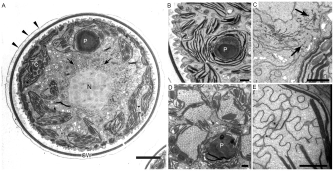
Figure 2. Transmission electron micrographs of green coccoid cells in H. pluvialis.
A. General ultrastructure. The cell wall is surrounded by extracellular matrix (arrowheads). Arrows indicate astaxanthin granules. B. Chloroplast and pyrenoid. C. High-magnification view of astaxanthin granules (arrows). D, E. One-layer thylakoids with a regular arrangement. C, chloroplast; CW, cell wall; N, nucleus; P, pyrenoid. Scale bars in A and B–E: 5 µm and 1 µm, respectively.

