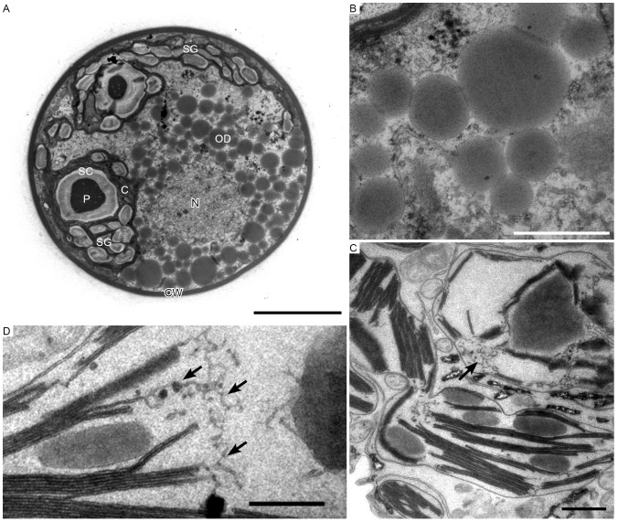
Figure 3. Transmission electron micrographs of intermediate H. pluvialis cells.
A. General ultrastructure. B. High-magnification view of astaxanthin oil droplets. C. Partial degradation of thylakoids (arrow). D. High-magnification view of thylakoid degradation (arrows). C, chloroplast; CW, cell wall; N, nucleus; OD; oil droplet; P, pyrenoid; SC, starch capsule; SG, starch grain. Scale bars in A and B–D: 5 µm and 1 µm, respectively.

