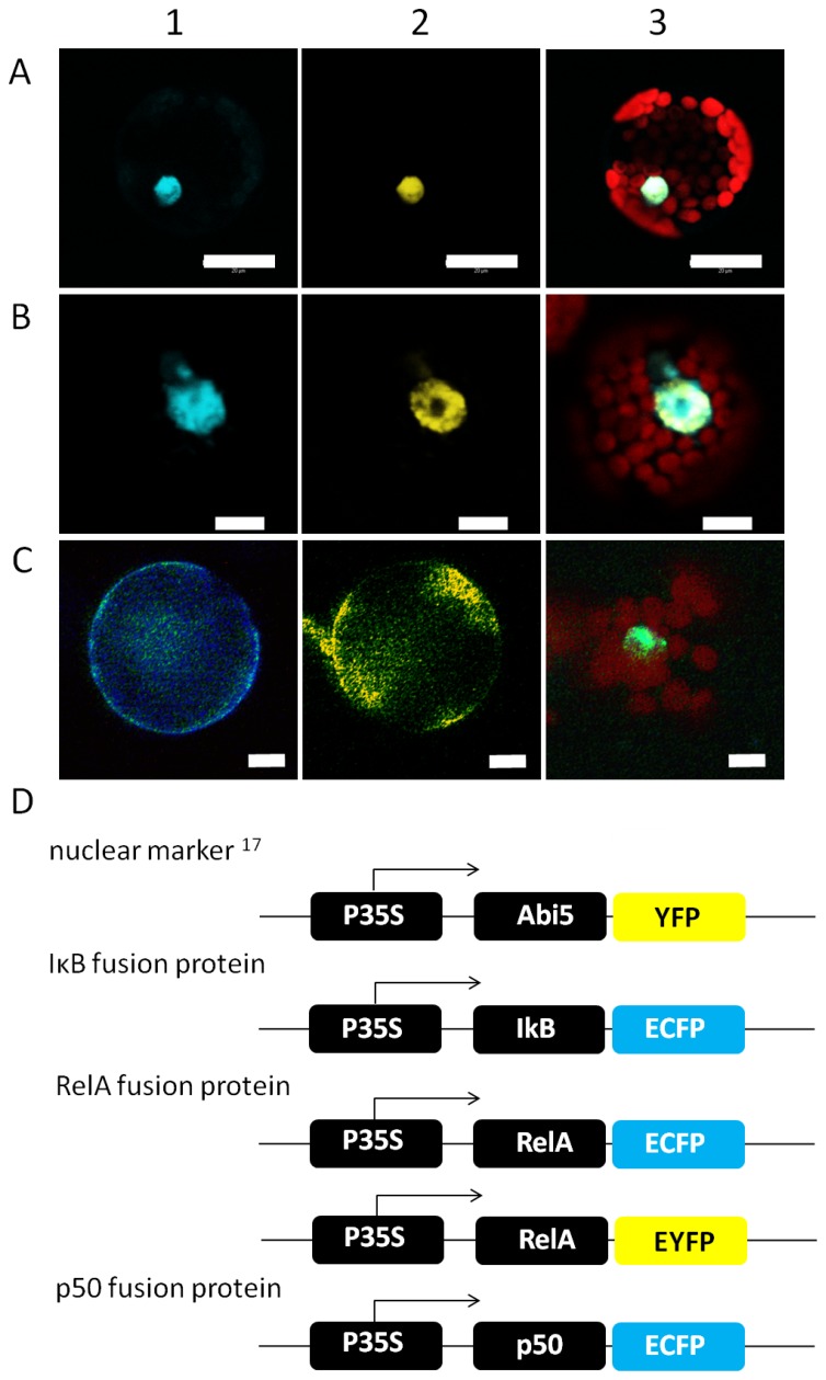
Figure 2. Subcellular Localization of p50, RelA and IκB in plant cells.
A, B) Co-localisation of the NF-κB subunits p50 (A) and RelA (B) with the plant transcription factor Abi5 (Lopez-Molina et al. 2003). The fusion proteins were expressed in Arabidopsis protoplasts and detected by confocal laser scanning microscopy. Abi5 was fused to YFP (A2, B2 yellow), the NF-κB subunits to ECFP (A1, B1 cyan). Plastid-derived autofluorescence is shown in red in the overlay images (A3, B3). C) Effect of IκB on the localisation of RelA in plant cells observed by fluorescence microscopy. The localisation of IκB is shown in cyan (C1), yellow staining has been chosen for the localization of RelA (C2) and the image (C3 shows Hoechst staining (green) of the nucleus as well as the chlorophyll autofluorescence in red. In (C) RelA and IκB are not co-localized with nuclear staining. Representative images are shown. Scale bars correspond to 20 (A) and 10 µm (B, C). D) Construct design of Abi5-YFP, IκB-ECFP, p50-ECFP and RelA-EYFP under control of the CaMV35S-promoter.

