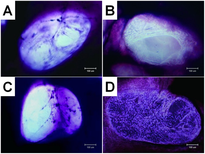
Figure 3. Photomicrographs (×100, methyl violet staining) of cell-scaffold constructs after in vitro culture for 12 d.
The number of attached cells and density of extracellular matrix (ECM) fibers in the interior of the scaffold are obvious different among four groups, with group B (B) > group D (D) > group A (A) > group C (C). Bar lengths are 100 um.

