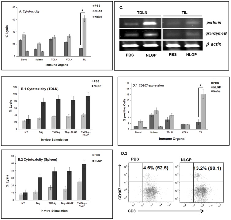
Figure 5. In vitro cytotoxicity of sarcoma by immune cells from NLGP-treated mice.
Mice were inoculated with Sarcoma 180 cells (1×10⁶ cells/mice) and after formation of palpable tumor, mice of experimental group were treated with NLGP once a week. Mice were sacrificed on day 21 to collect blood, spleen, TDLN, VDLN and tumor and MNCs were purified. MNCs are also purified from blood and spleen of naive mice. MNCs were co-cultured with sarcoma cells in 1∶10 ratios for 24 hrs and released LDH was measured (A). *p<0.001. MNCs from TDLN (B.1) and Spleen (B.2) of PBS- and NLGP- treated sarcoma bearing mice were cultured in vitro in presence of IL-2 for 4 days and further stimulated with tumor (sarcoma) antigens (5 µg/ml) for 2 days with or without NLGP. Antigens from tumor as well as its microenvironment were extracted from intra-peritoneally grown sarcoma (Tum-Ag) and from solid tumor (TME-Ag). Cytotoxic efficacy of activated T-cells was analyzed towards Sarcoma 180 cells by LDH release assay. C. Total RNA was isolated from MNCs of TDLN and tumor from PBS- and NLGP-treated mice on day 21 and cytotoxicity related genes (perforin and granzymeB) were analyzed at transcriptional level by RT-PCR. D. MNCs from all immune compartments were incubated with sarcoma cells to detect CD107a expression by intracellular flow cytometric staining. Data is presented in bar diagram (*p<0.001) (D.1) and a representative dot plot for CD107a expression from TILs is also shown (D.2).

