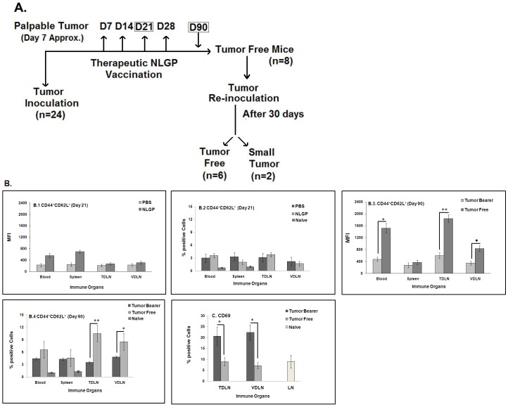
Figure 6. Memory response in sarcoma bearing mice after NLGP therapy.
A. A flow chart explaining the experimental design to understand the generation of memory response following NLGP therapy and re-inoculation with sarcoma in tumor free mice. Mice were inoculated with Sarcoma 180 cells and after formation of palpable tumor mice of the experimental group were treated with NLGP (25 µg) once a week for 4 weeks in total. A group of mice was sacrificed on day 21 to collect blood, spleen, TDLN and VDLN. MNCs were purified to stain with CD44 and CD62L antibodies, along with staining for CD8+ T cells, in comparison to cellular components obtained from PBS injected sarcoma bearing mice. Expression on blood, spleen and lymph node cells from naive mice was also studied. Both status of % positive cells (B.2) and MFI (B.1) are shown. Mice were inoculated with Sarcoma 180 cells and after the formation of palpable tumor, mice of experimental group were treated with NLGP (25 µg) once a week for 4 weeks in total. Tumor free mice were selected following 90 days of the initiation of therapy and re-inoculated with sarcoma. Blood, Spleen, TDLN and VDLN were isolated from representative mice of same group on day 90 and MNCs were purified to stain with CD44 and CD62L antibodies, in comparison to cellular components obtained from untreated sarcoma bearing mice. Both status of % positive cells (B.4) and MFI (B.3) are shown. In experiments described in B.1–B.4, CD8+ cells were first gated and CD44+CD62L+ cells within this population were then analyzed. *p<0.01; **p<0.001; • p<0.05. Percentage of CD69+ cells were analyzed within CD8+ cells from TDLN and VDLN on day 90 of identical experimental settings, mentioned in B. *p<0.01. Expression of CD69 on CD8+ T cells from lymph node of naive mice is also shown (C).

