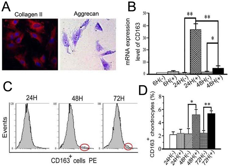
Figure 4. Exogenous TNF-α increased CD163 expression in primary chondrocytes from TMJ cartilage of 3-week old rats.
A: The primary cells isolated from TMJ cartilage of 3-week old rats were positive for type II collagen (Col-II) and aggrecan, as detected by immunofluorescence and toluidine blue, respectively (400× magnification). B: A time-course of induction of CD163 mRNA expression in primary cells isolated from TMJ cartilage and treated with 10 ng/ml of TNF-α. C–D: Flow cytometric analysis and graphical representation of the percentage of CD163+ cells within the primary cells isolated from TMJ cartilage and treated with 10 ng/ml of TNF-α.

