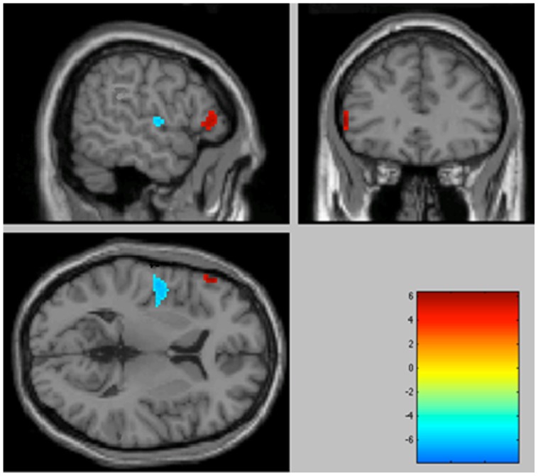
Figure 1. Significantly increased metabolic activity in the OCD patient group compared with healthy volunteers in the left inferior frontal gyrus (BA 45), associated with a decrease in activity in the left insula (BA 13) (p<0.001 uncorrected, colour bar represents t values).
Sagittal, coronal and transversal views in projection onto brain slices of a standard MRI (x/y/z coordinates according to Talairach atlas).

